Shoulder Arthroscopy
- drchiragthonse

- Dec 10, 2020
- 13 min read
Updated: Dec 11, 2020
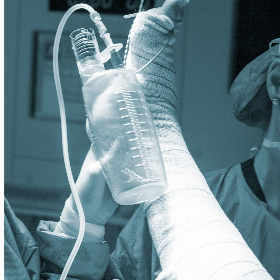
Shoulder arthroscopy is a surgery that uses a tiny camera called an arthroscope to examine or repair the tissues inside or around your shoulder joint. The arthroscope is inserted through a small cut (incision) in your skin.

Description
A rotator cuff is a group of muscles and their tendons that form a cuff over the shoulder joint. These muscles and tendons hold the arm in the shoulder joint, which also helps the shoulder move in different directions. The tendons in the rotator cuff can tear when they are overused or injured. You will likely receive general anaesthesia for this surgery, which means you will be asleep and unable to feel pain. Or, you may have regional anaesthesia. Your arm and shoulder area will be numbed. As a result, you do not feel any pain. If you receive regional anaesthesia, you will also be given medicine to make you very sleepy during the operation.
During the procedure, the surgeon:
Inserts the arthroscope into your shoulder through a small incision. The scope is connected to a video monitor in the operating room.
Inspects all the tissues of your shoulder joint and the area above the joint. These tissues include cartilage, bones, tendons, and ligaments.
Repairs any damaged tissues. To do this, your surgeon makes 1 to 3 more small incisions and inserts other instruments through them. A tear in a muscle, tendon, or cartilage is fixed. Any damaged tissue is removed. Your surgeon may do one or more of these procedures during your operation.
Rotator cuff repair:
The edges of the tendon are brought together. The tendon is attached to the bone with sutures.
Small rivets (called suture anchors) are often used to help attach the tendon to the bone.
The anchors can be made of metal or plastic. They do not need to be removed after surgery.
Surgery for impingement syndrome:
The damaged or inflamed tissue is cleaned out in the area above the shoulder joint.
A ligament called the coracoacromial ligament may be cut.
The underside of a bone called the acromion might be shaved. A bony growth (spur) on the bottom of the acromion often causes impingement syndrome. The spur can cause inflammation and pain in your shoulder.
Surgery for shoulder instability:
If you have a torn labrum, the surgeon will repair it. The labrum is the cartilage that lines the rim of the shoulder joint.
Ligaments that attach to this area will also be repaired.
The Bankart lesion is a tear on the labrum in the lower part of the shoulder joint.
A SLAP lesion involves the labrum and the ligament on the top part of the shoulder joint.
At the end of the surgery, the incisions will be closed with stitches and covered with a dressing (bandage). Most surgeons take pictures from the video monitor during the procedure to show you what they found and the repairs that were made. Your surgeon may need to do open surgery if there is a lot of damage. Open surgery means you will have a large incision so that the surgeon can get directly to your bones and tissues.
Why the Procedure is Performed
Arthroscopy may be recommended for these shoulder problems:
A torn or damaged cartilage ring (labrum) or ligaments
Shoulder instability, in which the shoulder joint is loose and slides around too much or becomes dislocated (slips out of the ball and socket joint)
A torn or damaged biceps tendon
A torn rotator cuff
A bone spur or inflammation around the rotator cuff
Inflammation or damaged lining of the joint, often caused by an illness, such as rheumatoid arthritis
Arthritis of the end of the clavicle (collarbone)
Loose tissue that needs to be removed
Shoulder impingement syndrome, to make more room for the shoulder to move around
Risks
Risks of anaesthesia and surgery in general are:
Allergic reactions to medicines
Breathing problems
Bleeding, blood clots, infection
Risks of shoulder arthroscopy are:
Shoulder stiffness
Failure of the surgery to relieve symptoms
The repair fails to heal
The weakness of the shoulder
Blood vessel or nerve injury
Damage to the cartilage of shoulder (chondrolysis)
Before the Procedure
Tell your health care provider what medicines you are taking, which includes medicines, supplements, or herbs you bought without a prescription. During the two weeks before your surgery:
You may be asked to stop taking blood thinners temporarily. These include aspirin, ibuprofen (Advil, Motrin), naproxen (Naprosyn, Aleve), and other medicines.
Ask your provider which medicines you should still take on the day of your surgery.
If you have diabetes, heart disease, or other medical conditions, your surgeon may ask you to see your doctor who treats you for these conditions.
Tell your provider if you have been drinking a lot of alcohol, more than 1 or 2 drinks a day.
If you smoke, try to stop. Ask your provider for help. Smoking can slow wound and bone healing.
Tell your doctor about any cold, flu, fever, herpes breakout, or other illness you may have before your surgery.
On the day of surgery:
Follow instructions about when to stop eating and drinking.
Take any drugs you're asked to take with a small sip of water.
Follow instructions on when to arrive at the hospital. Be sure to arrive on time.
After the Procedure
Follow any discharge and self-care instructions you are given. Recovery can take 1 to 6 months. You will probably have to wear a sling for the first week. If you had a lot of repairs done, you might have to wear the sling longer. You may take medicine to control your pain. When you can return to work or play sports will depend on what your surgery involved. It can range from 1 week to several months. Physical therapy may help you regain motion and strength in your shoulder. The length of treatment will depend on what was done during your surgery.
Prognosis
Arthroscopy often results in less pain and stiffness, fewer complications, a shorter (if any) hospital stay, and faster recovery than open surgery. If you had a repair, your body needs time to heal, even after arthroscopic surgery, just as you would need time to recover from open surgery. Because of this, your recovery time may still be long. Surgery to fix a cartilage tear is usually done to make the shoulder more stable. Many people recover fully, and their shoulder stays stable. But some people may still have shoulder instability after arthroscopic repair. Using arthroscopy for rotator cuff repairs or tendinitis usually relieves the pain, but you may not regain all of your strength.
LABRAL TEAR
Description
The labrum is a soft cartilage structure on the socket of the "ball and socket" joint (akaGH joint). It acts as a cushion between the two bones and serves as an attachment for the ligaments that stabilize the GH joint and the long head biceps tendon. The labrum can be torn from its attachment anywhere on the socket.
What are the causes?
Labral tears occur with acute (shoulder dislocations) or chronic, repetitive stresses to the GH joint. Athletes in collision sports, where the arm may be forced away from the body or with repetitive motion (baseball, volleyball, etc.) predispose the labrum to injury.
What are the symptoms?
Labral tears may cause different symptoms depending on the area of torn tissue. Labral tears in the front of the shoulder (anterior) are usually due to dislocations so that patients may experience pain and laxity in the shoulder. Labral tears in the back of the shoulder (posterior) are typically caused by repetitive stresses forcing the ball to the back of the socket. Patients may experience pain with stressful forces but not laxity in the shoulder. Labral tears in the top of the shoulder (superior) typically have pain and weakness, particularly with overhead activities like throwing. Pain may also be referred to the biceps, as its insertion on the labrum may have damage. Patients with labral tears may experience clicking in the shoulder.
How is it diagnosed?
Your surgeon will perform a thorough history and physical exam with X-rays. The shoulder will be moved through a range of motion and stressed in specific ways to elicit pain or feelings of laxity. X-rays may or may not show damage to the bones of the GH joint, particularly after an acute injury or dislocation. MRI (with contrast dye) helps evaluate the labrum, biceps and rotator cuff for damage.
How is it treated?
Non-operative
Labral tears in sedentary or older patients or the non-dominant extremity of an athlete may be successfully treated non-operatively. Physical therapy to strengthen the rotator cuff will help stabilize the humeral head in the socket and reduce pressure on the labrum. Anti-inflammatory medications, cryotherapy, activity modification or injection may be offered to treat pain.
Operative
Labral tears that cause pain or feelings of instability in the dominant extremity of athletes, and patients who have failed non-operative treatment require a minimally-invasive arthroscopic surgery to repair and reapproximate the tissue. Labral tears are sometimes more degenerative and do not need formal repair. In such cases, the damaged tissue is removed. If the labrum is not repairable, it is trimmed away. Labral tears which can be repaired are reattached to the socket. Young patients who have suffered shoulder dislocations should undergo operative repair, as the labrum and ligaments injured during a dislocation do not heal, which leaves the patient at risk for future dislocations and arthritis. Tears in the top part of the labrum (aka –SLAP tears) are treated identically, except when the biceps tendon is damaged. If the biceps attachment is severely compromised, the biceps is released from the socket and reattached outside the shoulder joint (biceps tenodesis) or released and not repaired outside of the joint (biceps tenotomy).
ROTATOR CUFF TEAR
Description
A rotator cuff is a group of four muscles that surround the GH joint. The muscles attach to the bone by thickened portions of non-elastic tissue called tendons. The rotator cuff is responsible for the majority of motion that occurs at the shoulder. Tears can happen in any of the four tendons. In order of commonality, the supraspinatus is most often injured, followed by the infraspinatus, subscapularis and teres minor. Tears in the tendons can either be partial or complete.
What are the causes?
Rotator cuff tears have multiple causes. Acute trauma, longstanding impingement syndrome (caused by abrasion on the acromion or bone spurs at the AC joint), repetitive overhead activities and degeneration in the older population are all standard.
What are the symptoms?
Rotator cuff tears cause pain, particularly on the side of the shoulder and at night, leading the patient to believe they slept wrong on their shoulder. Subacromial bursitis typically accompanies rotator cuff tears. Depending on the severity and location of the tear, loss of motion and strength can occur in a specific plane, which may be lifting the arm to the side, overhead or behind their back. Activities of daily living like reaching up to a cupboard, reaching for a wallet in a back pocket, brushing your air or fastening a bra may be severely impacted when a rotator cuff tear is present. A massive tear of more than one tendon may result in pseudoparalysis, an inability for the patient to move their arm away from their body. Pain may also radiate up the shoulder to the neck, as the patient attempts to move the shoulder using other muscles.
How is it diagnosed?
Your surgeon will perform a thorough history and physical exam, including X-rays. Exam findings will consist of loss of active range of motion (you move your arm), preserved passive range of motion (the surgeon moves your arm), weakness and pain with muscle testing. X-rays may or may not demonstrate acromion abnormalities or AC joint bone spurs that may be causing impingement of the rotator cuff. Superior or anterior movement of the humerus (ball) on X-ray is a sign of a chronic rotator cuff tear that may not be amenable to repair (aka-superior or anterior escape). MRI is useful to quantify the size, severity and age of the tear. For patients that cannot have an MRI, CT/" CAT" scan with contrast dye may help assess damage to the rotator cuff.
How is it treated?
Non-operative
Non-operative treatment can be attempted in patients with partial tears of the rotator cuff, or small tears with minimal symptoms. Physical therapy, anti-inflammatory medication, cryotherapy, activity modification or injections into space just above the rotator cuff may alleviate pain and inflammation. Patients whose pain does not resolve with non-operative treatment should discuss surgical treatment options with their surgeon.
Operative
Partial tears that fail to improve with non-operative treatment can be treated with a minimally-invasive arthroscopic procedure. Depending on the tear pattern and quality of the tissue, the partial tear can be "cleaned-up" (aka – debrided) or repaired. If the tear is due to an acromion abnormality or bone spur from AC joint arthritis, a subacromial decompression can be performed to eliminate the cause of the tear. Tears in which the tendon has detached from the bone should be surgically repaired, as they cannot heal on their own. Your surgeon may perform a minimally-invasive arthroscopic procedure or through an open incision. Following rotator cuff repair, your surgeon will recommend a sling to protect the repair while it heals, and start physical therapy to regain motion and strength in the shoulder. The length of time in a sling and the start of physical therapy are based on the type of tear, tissue quality and ability to heal, among other factors. In cases where the rotator cuff cannot be repaired, the surgeon can choose to debride the torn tendon and attempt a partial rotator cuff repair for pain relief and function. A minimally-invasive arthroscopic superior capsular reconstruction (SCR) can be completed in a young active patient without arthritic changes and in the presence of an irreparable rotator cuff tear for pain relief and potentially some restoration of motion. Finally, a reverse total shoulder replacement can be performed for pain relief and potentially, some restoration of motion if soft tissue repair is not possible.
PROXIMAL BICEPS RUPTURE
Description
The biceps muscle has two origin points around the shoulder: the long head starts on the glenoid and the short head on the coracoid. The long head is more commonly injured/inflamed as it exits the "ball and socket" joint near the rotator cuff in the front of the shoulder. Proximal biceps rupture is the spontaneous tearing of the long head of the biceps from its origin on the glenoid.
What are the causes?
Proximal biceps rupture typically occurs with trauma, falls or sudden lifting. The tendon may be predisposed to rupture if it is chronically inflamed (tendinitis) or with an associated rotator cuff tear.
What are the symptoms?
Proximal biceps tendon rupture causes immediate pain, swelling and bruising in the front of the shoulder. With time, bruising travels down the front of the arm towards the muscle. Simultaneously, a bulge in the front of the arm appears and the muscle contracts, which is known as a "Popeye" deformity. Muscle spasm and weakness with flexing the elbow and rotating the wrist may be noticed with proximal biceps rupture.
How is it diagnosed?
Your surgeon will perform a thorough history and physical exam, often including X-rays. Proximal biceps rupture is diagnosed in the office. An MRI or ultrasound may help determine if there is damage to the rotator cuff. In either case, the long head of the biceps will not appear at its origin on the glenoid and will not occupy the normal space on the front of the shoulder (bicipital groove).
How is it treated?
Non-operative
Non-operative treatment of proximal biceps rupture is common. Physical therapy, anti-inflammatory medication, cryotherapy and activity modification early after the injury help reduce pain, inflammation and muscle spasm. Over time, patients may have significant pain relief if they suffered from chronic biceps tendinitis before the injury. Most patients will not see a substantial decrease in elbow or wrist function since the short head of the biceps is still intact. The "Popeye" deformity is permanent, but the cosmetic deformity does not affect overall function.
Operative
Surgical intervention for proximal biceps rupture is limited to athletes, particularly overhead athletes, patients with manual labour occupations that include repetitive elbow and wrist motion (electricians, carpenters, etc.) as fatigue and muscle spasm may occur and patients that do not want the "Popeye" deformity. In these cases, your surgeon will perform a biceps tenodesis, reattaching the tendon to a site along the arm bone. The reattachment is not performed at the site of injury at the tendon origin.
ROTATOR CUFF ARTHROPATHY
Description
A rotator cuff is a group of 4 muscles that help move the shoulder. The muscles attach to the bones around the ball-and-socket joint by a thick, non-elastic tissue called a tendon. Tendons can tear by an acute injury or degenerate over time. Tears can occur in any of the four tendons. In order of commonality, the supraspinatus is most often injured, followed by the infraspinatus, subscapularis and teres minor. Rotator cuff arthropathy is defined as arthritis as the result of the tearing of the rotator cuff tendons.
What are the causes?
Rotator cuff tears have multiple causes. Acute trauma, longstanding impingement syndrome (caused by abrasion on the acromion or bone spurs at the AC joint), repetitive overhead activities and degeneration in the older population are all common. When the tendons are detached from the bone, the shoulder becomes dysfunctional. Pain is associated with the motion of the arm, and movement eventually decreases if the tendons are not repaired back to the bone.
What are the symptoms?
Rotator cuff tears cause pain, particularly on the side of the shoulder and at night, leading the patient to believe they slept wrong on their shoulder. Subacromial bursitis typically accompanies rotator cuff tears. Depending on the severity and location of the tear, loss of motion and strength can occur in a specific plane, which may be lifting the arm to the side, overhead or behind their back. Activities of daily living like reaching up to a cupboard, going for a wallet in a back pocket, brushing your hair or fastening a bra may be severely impacted when a rotator cuff tear is present. A massive tear of more than one tendon may result in pseudoparalysis, an inability for the patient to move their arm away from their body. Pain may also radiate up the shoulder to the neck, as the patient attempts to move the shoulder using other muscles. If the tendons remain torn for an extended period, the ball does not stay centred in the joints, causing wear and tear to one or both sides of the ball-and-socket joint.
How is it diagnosed?
Your surgeon will perform a thorough history and physical exam, including X-rays. Exam findings will consist of loss of active range of motion (you move your arm), preserved passive range of motion (the surgeon moves your arm), weakness and pain with muscle testing. X-rays may or may not demonstrate acromion abnormalities or AC joint bone spurs that may be causing impingement of the rotator cuff. Superior or anterior movement of the humerus (ball) on X-ray is a sign of a chronic rotator cuff tear that may not be amenable to repair (also known as superior or anterior escape). MRI is useful to quantify the size, severity and age of the tear. For patients who cannot have an MRI, CT/" CAT" scan with contrast dye may help assess damage to the rotator cuff.
How is it treated?
Non-operative
The extent of arthritis, long term damage to the tendons and muscles, the function of the shoulder, amount of pain and patient factors (age, health issues like diabetes or seizure disorder, tobacco use and activity level) all influence the treatment of rotator cuff arthropathy. Non-operative treatment can be attempted but once the diagnosis of rotator cuff arthropathy is made, is not usually helpful. Physical therapy, anti-inflammatory medication, cryotherapy, activity modification or injections into space just above the rotator cuff may alleviate pain and inflammation. Patients whose pain does not resolve with non-operative treatment should discuss surgical treatment options with their surgeon.
Operative
Patients who have a chronic, irreparable tear in the rotator cuff but have minimal to no arthritis, several minimally invasive arthroscopic procedures can be performed for pain relief. An arthroscopic CAM procedure, also known as debridement or "cleanout", can remove inflammatory tissue, remove bone spurs and treat biceps tendon injury/inflammation. An arthroscopic superior capsular reconstruction (SCR) uses donor tissue to realign the ball-and-socket joint and restore some of the mechanics that are affected by chronic rotator cuff tendon tears. Both of these procedures may decrease pain, but the restoration of function is patient dependent. For more advanced rotator cuff arthropathy with moderate to severe arthritis, reverse total shoulder arthroplasty (rTSA) can be performed for pain relief and restoration of some function. In rTSA, your surgeon can remove the arthritic areas of the ball-and-socket joint and replace them with metal and plastic components. In this procedure, the ball-and-socket elements are switched, so that the ball becomes a socket and the socket becomes a ball, allowing the shoulder to move pain-free and using other muscles instead of the rotator cuff. Your surgeon will discuss all of your options based on the severity of damage in your shoulder.


Comments