Arthroscopy
- drchiragthonse

- Dec 11, 2020
- 13 min read
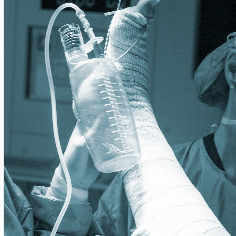
Arthroscopic Surgery is a procedure that allows surgeons to see, diagnose, and treat problems inside a joint. The procedure, also called an Arthroscopy, requires only small incisions and is guided by a miniature viewing instrument or scope. Before arthroscopy existed, surgeons made large incisions that affected the surrounding joint structures and tissues. They had to open the joint to view it and perform surgery. The traditional surgery method carries a higher risk of infection and requires a longer time for recovery.
In contrast, arthroscopy is less invasive. It has a decreased risk of infection and a shorter
recovery period. Today, arthroscopic surgery is one of the most common orthopaedic
procedures.
Why it’s done?
Doctors use arthroscopy to help diagnose and treat a variety of joint conditions, most commonly those affecting the:
● Knee
● Shoulder
● Elbow
● Ankle
● Hip
● Wrist
Knee Arthroscopy

● ACL tear/rupture: ACL reconstruction is now a commonly performed arthroscopic procedure.
● Meniscal tear/deficiency: The meniscus is a vital structure and can be repaired arthroscopically. If irreparable, surgeons remove the tear and stabilize the remnant.
● PCL rupture/rupture: PCL reconstruction can also be performed arthroscopically.
● Collateral Ligament tear
● Chondral defects
● Articular cartilage damage (early arthritis): If caught early, articular cartilage damage can be repaired and stabilized. Otherwise, surgeons remove the unstable flaps and prepare the underlying bone to encourage new cartilage to form, a process known as ‘micro-fracture’.
● Synovitis: Inflamed tissue around the knee can be removed (i.e., synovectomy).
● Loose body: Bone, cartilage or any other loose tissue can be removed to prevent ongoing damage to the knee.
● Biopsy: Tissue specimens can be taken to aid in the diagnosis of certain conditions.
● Washouts: For the treatment of knee infections
ACL

Description
The anterior cruciate ligament (ACL) is the main stabilizing ligament on the inside of the knee. Its primary function is to prevent the tibia (shin bone) from sliding forward and rotating on the femur (thigh bone). Tears/ruptures of the ligament result in knee instability.
What are the causes?
ACL tears are typically caused by twisting or hyperextension injuries. Sports activity like pivoting or sudden deceleration when running and falls during skiing are considered non-contact causes of ACL tears. Direct trauma to the back or side of the knee during collision sports is regarded as a contact injury to the ACL.
What are the symptoms?
ACL tears cause immediate pain and often swelling. You may feel something “pop” inside the knee. An initial inability to bear weight on the leg may subside and walk may be possible after several minutes. The knee may feel loose or that it is going to “give out” and return to sport is impossible. Over time, swelling will increase, and motion may be lost.
How is it diagnosed?
Your surgeon will perform a thorough history and physical exam with X-rays. On exam, swelling and loss of motion and strength are present. Your surgeon will perform manoeuvres to check the stability of all the knee ligaments and the meniscus. An MRI is helpful to confirm the diagnosis, showing the ACL tear. The type of tear (partial, complete, avulsion from either the tibia or femur) can be defined, which may assist in surgical planning. The MRI may also show bruising bone secondary to the injury.
How is it treated?
Non-operative
ACL tears do not heal. Some patients elect not to have reconstruction surgery. Non-operative treatment increases the risk of “wear and tear” arthritis and meniscus tears because of the instability in the joint. Non-operative treatment consisting of anti-inflammatory medication, physical therapy, cryotherapy and activity modification may be prescribed before surgery to decrease the swelling, regain motion and strength, as research has demonstrated that surgery is less complicated and patients have better outcomes. Non-operative treatment in a surgical patient may be skipped if other injuries to the meniscus and cartilage are present and need to be repaired immediately.
Operative
Operative management of ACL tears depends on the type of tear. ACL repair may be indicated in patients where the ACL is torn off the wall of the femur (thigh bone) or tibia (shin bone. ACL repair is accomplished through a minimally-invasive arthroscopic procedure and sewed back into place and fixed with screws or buttons. The repair may also be supplemented with high-strength suture.
If formal reconstruction is required, a new ACL graft will be fixed in place of the original ligament. A technique for graft placement and graft choice is a shared decision between you and your surgeon. Most techniques are performed through a minimally-invasive arthroscopic procedure. The graft can be taken from around your knee or from a donor. Postoperative rehabilitation, return to daily activities and return to sport depends on the technique and graft chosen, and is at your surgeon’s discretion.
PCL

Description
The posterior cruciate ligament (PCL) is the other main stabilizing ligament on the inside of the knee. Its primary function is to prevent the tibia (shin bone) from sliding backwards and rotating on the femur (thigh bone). Tears/ruptures of the ligament result in knee instability. PCL tear is less common than an ACL tear.
What are the causes?
PCL tears are typically caused by trauma or a fall on the knee. A direct posterior/backward force on the tibia commonly seen in collision sports or the knee hitting the dashboard in a motor vehicle accident will cause a PCL tear.
What are the symptoms?
PCL tears cause immediate pain and often swelling. You may feel something “pop” inside the knee. An initial inability to bear weight on the leg may subside and walk may be possible after several minutes. The knee may feel loose or that it is going to “give out” and immediate return to sport is impossible. Over time, swelling will increase, and motion may be lost. Unlike ACL tears, some patients, even athletes, can return to sport with partial PCL tears (albeit in a knee brace) and never require surgery.
How is it diagnosed?
Your surgeon will perform a thorough history and physical exam with X-rays. On exam, swelling and loss of motion and strength are present. Your surgeon will perform manoeuvres to check the stability of all the knee ligaments and the meniscus. An MRI is helpful to confirm the diagnosis, showing the PCL tear. The type of tear (partial, complete, avulsion from either the tibia or femur) can be defined, which may assist in treatment planning. The MRI may also show bruising bone secondary to the injury.
How is it treated?
Non-operative
PCL tears do not heal. However, some patients may be able to return to regular activity
depending on the type and severity of the tear. Non-operative treatment consisting of
anti-inflammatory medication, physical therapy, cryotherapy and activity modification
may be prescribed to decrease the swelling, regain motion and strength. A brace may
be prescribed to return to sports activities. If symptoms persist (pain, instability),
reconstruction surgery may be recommended by your surgeon.
Operative
Operative management of PCL tears depends on the type of tear. PCL repair may be
indicated in patients where the PCL is torn off the wall of the femur (thigh bone) or tibia
(shin bone. PCL repair is accomplished through a minimally-invasive arthroscopic
procedure and sewed back into place and fixed with screws or buttons. The repair may
also be supplemented with high-strength suture.
If formal reconstruction is required, a new PCL graft will be fixed in place of the original
ligament. A technique for graft placement and graft choice is a shared decision
between you and your surgeon. Most techniques are performed through a
minimally-invasive arthroscopic procedure. The graft can be taken from around your
knee or from a donor. Postoperative rehabilitation, return to daily activities and return
to sport depends on the technique and graft chosen, and is at your surgeon’s
discretion.
MENISCAL TEAR

Description
The meniscus is cartilage that acts as a shock absorber between the femur (thigh bone) and tibia (shin bone). Each knee has two distinct menisci: the medial (an inner aspect of the knee) and lateral (outer part of the knee). Medial meniscus tears are more common in general, and lateral meniscus tears are more common when the ACL is injured. Injuries to the meniscus may lead to eventual degenerative changes in the knee (aka – arthritis).
What are the causes?
The meniscus can be injured in several ways. Acute meniscus tears result from a sudden twisting or pivoting manoeuvre. Acute meniscus tears are associated with ACL injuries. The meniscus can also undergo degeneration as patient age increases. The degenerative meniscus is susceptible to tearing with minimal trauma (i.e.-twisting the knee getting into the car).
What are the symptoms?
Meniscus tears, in the acute setting, cause immediate pain over the specific meniscus, potentially swelling and bruising and loss of motion and strength. The patient may feel a clicking or catching with walking and increased pain with twisting on the affected foot. If the meniscus tears and gets stuck out of place, the knee may feel locked (aka -bucket handle meniscus).
How is it diagnosed?
Your surgeon will perform a thorough history and physical exam with X-rays. On exam, swelling and loss of motion and strength are present. The knee is painful to touch over the affected meniscus. Your surgeon may perform provocative manoeuvres to test each meniscus, resulting in pain and clicking if the meniscus is torn. X-rays are usually standard. MRI is helpful to confirm the diagnosis and characterize the tear for surgical planning. Other injuries can also be identified on the MRI.
How is it treated?
Non-operative
Some meniscus tears are treated successfully without surgery. Your surgeon may prescribe anti-inflammatory medication, physical therapy, cryotherapy and activity modification to reduce pain and inflammation, as well as strengthen the muscles around the knee to decrease the force transmitted to the meniscus. Your surgeon may offer you an injection. Patients with persistent symptoms (pain, clicking, etc.) may benefit from surgery. Bucket handle meniscus tears are not treated non-operatively and require surgery.
Operative
Meniscus tears can be treated in most cases with minimally-invasive arthroscopic surgery. Depending on the size and type of tear, as well as the quality of the torn tissue, your surgeon may choose to remove the torn meniscus or repair it with a series of sutures. Removing large portions of the meniscus will lead to expedited degeneration of the joint cartilage (aka- arthritis). Postoperative rehabilitation is at your surgeon’s discretion.
MCL REPAIR

Description
The medial collateral ligament (MCL) is the main stabilizing ligament on the inner aspect of the knee. Its primary function is to prevent the knee from buckling inward/knock-knee (valgus motion). Tears/ruptures of the ligament result in knee instability.
What are the causes?
MCL tears are typically caused by trauma. A direct force to the outside of the knee stresses the ligament, which naturally occurs in collision sports like football. Overuse injuries in sports/occupations that require repetitive falling to the knees and standing up quickly can also lead to micro-tears of the ligament.
What are the symptoms?
MCL tears cause immediate pain and often swelling. You may feel something “pop” on the inside aspect of the knee. Pain is centralized over the ligament (an inside element of the knee). Walking after the injury may be possible, but the knee may feel like it’s going to “give in” depending on the severity of the tear. The MCL is attached to the underlying meniscus. Damage to the meniscus at the time of injury may cause clicking or locking of the knee.
How is it diagnosed?
Your surgeon will perform a thorough history and physical exam with X-rays. On exam, swelling and loss of motion and strength are present. Your surgeon will perform manoeuvres to check the stability of all the knee ligaments and the meniscus. An MRI is helpful to confirm the diagnosis, showing the MCL tear. The type of tear (partial, complete, avulsion from either the tibia or femur) can be defined, which may assist in treatment planning. The MRI may also show bruising bone secondary to the injury.
How is it treated?
Non-operative
Almost all minor MCL tears can be treated non-operatively. Non-operative treatment consisting of bracing, anti-inflammatory medication, physical therapy, cryotherapy and activity modification may be prescribed to decrease the swelling, regain motion and strength. Most patients may be able to return to regular activity without surgery, depending on the type and severity of the tear. A brace may be prescribed to return to sports activities. If symptoms persist (pain, instability), reconstruction surgery may be recommended by your surgeon.
Operative
Operative management of MCL tears depends on the type of tear. MCL repair may be indicated in patients where the MCL is torn off the wall of the femur (thigh bone) or tibia (shin bone). MCL repair is accomplished through a series of small incisions and sewed back into place and fixed with screws or buttons. The repair may also be supplemented with high-strength suture.
If formal reconstruction is required, a new MCL graft will be fixed in place of the
original ligament. A technique for graft placement and graft choice is a shared decision
between you and your surgeon. Most procedures are performed through a
minimally-invasive incision. The graft can be taken from around your knee or from a
donor. Postoperative rehabilitation, return to daily activities and return to sport
depends on the technique and graft chosen, and is at your surgeon’s discretion.
LCL

Description
The lateral collateral ligament (LCL) is the main stabilizing ligament on the outer aspect of the knee. Its primary function is to prevent the knee from giving way outward (varus motion). Tears/ruptures of the ligament result in knee instability.
What are the causes?
Isolated LCL tears are uncommon. They typically occur from trauma. A direct force to the inside of the knee stresses the ligament, which naturally occurs in collision sports like football. LCL tears are also seen in high-energy trauma like motor vehicle accidents. They are accompanied by tears in the other ligaments and tendons on the outside of the knee (aka – posterolateral corner injury and knee dislocation).
What are the symptoms?
LCL tears cause immediate pain and often swelling. You may feel something "pop" on the outer aspect of the knee. Pain is centralized over the ligament (an outside part of the knee). Walking after the injury may be possible, but the knee may feel like it's going to "give out" depending on the severity of the tear.
How is it diagnosed?
Your surgeon will perform a thorough history and physical exam with X-rays. On exam, swelling and loss of motion and strength are present. Your surgeon will perform manoeuvres to check the stability of all the knee ligaments and the meniscus. An MRI is helpful to confirm the diagnosis, showing the LCL tear. The type of tear (partial, complete, avulsion from either the tibia or femur) can be defined, which may assist in treatment planning. The MRI may also show bruising bone secondary to the injury.
How is it treated?
Non-operative
Almost all minor LCL tears can be treated non-operatively. Non-operative treatment consisting of bracing, anti-inflammatory medication, physical therapy, cryotherapy and activity modification may be prescribed to decrease the swelling, regain motion and strength. Most patients may be able to return to regular activity without surgery, depending on the type and severity of the tear. A brace may be prescribed to return to sports activities. If symptoms persist (pain, instability), reconstruction surgery may be recommended by your surgeon. If other structures are damaged (i.e., Posterolateral Corner), surgery is recommended to reconstruct the knee.
Operative
Operative management of LCL tears depends on the type of tear. LCL repair may be
indicated in patients where the LCL is torn off the wall of the femur (thigh bone) or tibia (shin bone). LCL repair is accomplished through a series of small incisions and sewed back into place and fixed with screws or buttons. The repair may also be supplemented with high-strength suture.
If formal reconstruction is required, a new LCL graft will be fixed in place of the original
ligament. A technique for graft placement and graft choice is a shared decision between you and your surgeon. Most procedures are performed through a minimally-invasive incision. The graft can be taken from around your knee or from a donor. Postoperative rehabilitation, return to daily activities and return to sport depends on the technique and graft chosen, and is at your surgeon's discretion.
CHONDRAL DEFECTS

Description
Osteochondritis Dissecans (OCD) is a condition in which fragments of joint cartilage become separated from the bone. These fragments may peel from the bone and remain intact or completely separate from the bone, forming loose bodies that float in the joint. These fragments usually originate from the femoral (thigh bone) side of the joint but can create from any compartment.
What are the causes?
It is currently thought that most OCD lesions occur as a result of a traumatic injury that occurred in the patient's past. They can also occur in the athlete as a result of overuse. Some lesions do not have an identifiable cause.
What are the symptoms?
OCD causes pain at a specific area in the knee. Patients will complain of clicking and locking in the knee, mainly if the fragment has separated and is floating around the joint. Patients are often unable to flex and extend their knee fully.
How is it diagnosed?
Your surgeon will perform a thorough history and physical exam, which typically includes X-rays. Your surgeon will evaluate the range of motion. The origin of the fragment may be tender to the touch. X-rays may or may not identify OCD. MRI is standard for OCD diagnosis. Fluid seen behind the fragment on MRI is an indicator of OCD.
How is it treated?
Non-operative
Stable OCD lesions, those not likely to displace, are treated with anti-inflammatory medication, cryotherapy, activity modification and observation.
Operative
A minimally-invasive arthroscopic procedure evaluates unstable OCD lesions.
Sometimes the fragment can be repaired with small screws/tacks if it is still attached to
the bone. If the fragment has separated from the bone and cannot be repaired, it will be removed, and a cartilage regeneration or replacement procedure may be performed in the lesion. A microfracture procedure can be performed in the lesion to allow blood to soak the lesion. A cartilage graft, typically from a donor, can then be placed at the site. Over time, new cartilage will be formed over the lesion. Alternatively, your surgeon may choose to take a piece of cartilage from one area of the knee where it is not necessary and place it into the defect (autograft cartilage transplant procedure).
ARTHRITIS

Description
Arthritis is the inflammation of a joint. The knee can be divided into three compartments: medial (inside), lateral (outside) and patellofemoral (front). Arthritis can be present in one, two or three compartments. Over time, the loss of the smooth covering on the ends of bones (aka - articular cartilage) causes pain and stiffness, which can lead to pain with motion or at rest, swelling, clicking or grinding and a loss of strength. When the cartilage is damaged or decreased, the bones rub together during joint motion, resulting in "bone-on-bone" arthritis. When arthritis becomes severe, inflammation occurs around the joint and extra bone is formed in an attempt to protect the joint, resulting in limited motion and strength.
What are the causes?
The primary cause of arthritis is osteoarthritis (aka – "wear and tear" arthritis). Trauma
and other illnesses like rheumatoid arthritis, systemic lupus, septic arthritis and psoriasis can result in degeneration of a joint, leading to symptoms of pain and lack of motion.
What are the symptoms?
Arthritis of the knee causes pain, swelling, stiffness and loss of strength. Pain can be isolated to the medial, lateral or patellofemoral aspects of the joint or be generalized discomfort around the knee. Pain and swelling in the back of the knee may be from a Baker's Cyst, an area of the fluid collection that is caused by arthritis. A 'grinding', 'clicking' or 'locking' sensation may be felt. Loss of motion can become severe, and the patient may have trouble performing tasks, such as walking long distances. Patients who have arthritis of the patellofemoral joint will often complain of 'giving way' or buckling of the knee. Patients with patellofemoral arthritis have trouble using stairs, squatting, or standing after prolonged sitting.
How is it diagnosed?
Your surgeon will perform a thorough history and physical exam, which typically includes X-rays. Your surgeon will evaluate the range of motion, stability of the ligament and strength of the muscles surrounding the knee. X-rays may demonstrate decreasing space between the bones (joint space narrowing) and bone spurs (osteophytes) in areas of arthritis. MRI may be helpful to determine if other areas of joint cartilage or the meniscus have damage.
How is it treated?
Non-operative
Knee arthritis can be treated with physical therapy to strengthen the muscles that support the joint. The stronger the supporting muscles, the less the body will need to rely on bony architecture to stabilize the joint, which will lead to less stress across the arthritic area. Your surgeon may prescribe anti-inflammatory medication or offer an injection to reduce the inflammation. Certain nutritional supplements may be beneficial to decrease pain and inflammation.
Operative
When non-operative treatment does not relieve symptoms, your surgeon may suggest
surgery. Three surgical options are available for knee arthritis.
Minimally-invasive arthroscopy of the knee, or a 'knee scope', may be beneficial to "clean-out" the knee. Although not a cure, this procedure may provide relief in patients suffering mechanical symptoms, such as catching and locking. The entire knee joint, including joint cartilage, meniscus and ligaments, can be evaluated during arthroscopy.
The definitive treatment for knee arthritis is joint replacement surgery. Your surgeon will
resurface the ends of the bone where the cartilage has worn away, with metal and
plastic implants. If the arthritis is localized to a single or two compartments (medial,
lateral or patellofemoral), your surgeon will replace only the areas that are affected
(unicompartmental or patellofemoral replacement). If the arthritis is present in all three
compartments, a total knee replacement is required to alleviate symptoms.


Comments